
Abstract
Background
Septic arthritis is a potentially limb or life-threatening joint infection that requires prompt recognition and intervention to reduce morbidity and mortality. While intra-articular joint injections are commonly performed for osteoarthritis and other arthropathies, they carry a rare but significant risk of iatrogenic infection, particularly when performed in the presence of unrecognized joint or periarticular infection.
Case Presentation
We report a case of a 52-year-old female with a history of traumatic brain injury and chronic right knee pain who developed severe knee swelling, pain, and systemic symptoms following an intra-articular corticosteroid injection performed without ultrasound evaluation/guidance. Post-procedure, she presented with fever, elevated inflammatory markers, and purulent knee effusion. Operative washout revealed a purulent tract extending from the knee joint capsule to the lateral thigh. Cultures from joint aspiration and intraoperative samples grew Streptococcus dysgalactiae. Blood cultures were negative. The patient reported frequent cat scratches to the affected knee and described an unusual sensation of the injection needle tracking laterally during the joint injection. She was treated with surgical drainage and a four-week course of amoxicillin, with full clinical recovery.
Discussion
This case highlights an unusual presentation of septic arthritis with extra-capsular extension likely due to iatrogenic needle tracking during joint injection. The causative organism, S. dysgalactiae, is an uncommon pathogen in septic arthritis, and the presumed source was contiguous spread from untreated cellulitis, possibly related to cat scratches/bites. The absence of predisposing comorbidities and negative blood cultures further support a local rather than hematogenous source. The case emphasizes the importance of thorough clinical evaluation and consideration of infection prior to joint injections, as well as the potential benefits of ultrasound guidance to minimize procedural complications.
Conclusion
Careful history, physical examination, and appropriate imaging are essential prior to joint interventions to avoid iatrogenic complications. This case illustrates the rare but serious risk of extra-capsular extension of septic arthritis following intra-articular injection and emphasizes the need for vigilance in identifying underlying infection before proceeding with invasive procedures.
Author Contributions
Academic Editor: Ian James Martins, Principal Research Fellow Edith Cowan University
Checked for plagiarism: Yes
Review by: Single-blind
Copyright © 2025 Nawaf Al-saeed, et al
 This is an open-access article distributed under the terms of the Creative Commons Attribution License, which permits unrestricted use, distribution, and reproduction in any medium, provided the original author and source are credited.
Competing interests
The authors have declared that no competing interests exist.
Citation:
Introduction
Septic arthritis is a serious medical condition characterized by the infection of synovial fluid, primarily by bacteria. The incidence of septic arthritis ranges from 4-29 per 100,000 person-years with increased risk with advanced age, lower socioeconomic status, and immunosuppression1. Patients with this condition mainly present with mono-arthritis, fever and elevated inflammatory markers. Prompt diagnosis of septic arthritis is crucial, as the condition is associated with significant morbidity and mortality2. Management typically involves arthrocentesis and empiric antibiotic therapy, with surgical drainage indicated in some cases2. In adults aged 16 years or older, the most common cause of septic arthritis is Staphylococcus aureus as well as Neisseria gonorrhea3. Septic arthritis predominantly affects the knee, hip, ankle, wrist and shoulder joints in this population4.
Joint injections are a common procedure in the outpatient sport medicine and orthopedic setting. The procedure consists of injecting the targeted joint with both steroids and local anesthetic. The anesthetic often reduces post traumatic effect of injecting the joint and often acts as a short-term pain reliever until the steroids become effective. Steroids play a therapeutic role in reducing inflammation in the joint space and provide long lasting relief.5 Most common indications for joint injections include osteoarthritis, rheumatoid arthritis, gout, and adhesive capsulitis5. Other conditions may include tendinopathy, bursitis, carpal tunnel syndrome, trigger finger and many other conditions6. Furthermore, ultrasound has been utilized more to guide the needle in injections in joint spaces. Studies have consistently shown that ultrasound needle guidance usually results in more accurate joint injections, which often minimizes the morbidity associated with the procedure 7, 8. They have also been found to be more accurate and more efficacious compared to landmark guided injections.9Contraindications to joint injections include infection of the joint space or cellulitis in the skin overlying the joint10. Often times, adverse events of joint injections include infection, allergic reaction, systemic steroid effects, bleeding or ligament/tendon injury5. A concerning, but rare, side-effect of intra-articular injections also includes iatrogenic septic arthritis which has been estimated to occur in or less than 0.005% of joint injections.
To summarize, septic arthritis and joint injections are both important in the sports medicine setting. Septic arthritis is a serious joint infection requiring quick diagnosis and treatment to avoid possible complications. Joint injections, while generally safe, can have potential adverse effects, including the risk of introducing infection and it is critical to avoid joint injections during an active infection. Thus, careful consideration of the risks and benefits is necessary for both conditions, with ultrasound guidance being beneficial in both evaluation of the joint, as well as improving the safety and accuracy of joint injections.
In this article, we discuss a case of a patient who had a complicated course of septic arthritis of the knee as a complication of an intra-articular injection performed without prior ultrasonography nor ultrasound guidance which may have prevented this undesired outcome. Thus, highlighting the importance of ultrasound utilization prior and during intra-articular joint injections.
Clinical Course
52-year-old female known to have traumatic brain injury (TBI) since the 1980’s with associated tremors and no other medical comorbidities had been complaining of right sided knee pain for multiple years. She notes that due to the tremors from her TBI, she had been deviating a lot of weight over her right side. However, for 6 months, the patient had been complaining of worse than usual right knee pain, for which she had been referred to the sports medicine clinic by her primary care physician. In the clinic, intra-articular joint injection (IJI) without ultrasound guidance was discussed and performed. 6 days following the joint injection, she had noted significant worsening of her knee pain with associated swelling and systemic symptoms including fevers and night sweats. She had presented to the emergency department. Lab work revealed a C-reactive peptide levels of 467.3 mg/L, a white blood cell count of 15.2x10³ u/L and erythrocyte sedimentation rate of mm/hr. Arthrocentesis was performed with purulent material extracted, negative crystals, and notable for 175,0000 nucleated cells with 98% polymorphonucleated cells. She was started on linezolid 600 mg twice daily and taken for an operative knee washout. During the operation, a tract was noted to extend from the joint capsule to the lateral aspect of the thigh. External pressure to the lateral thigh was used to express purulent material through the tract, then an incision and drainage was performed to ensure complete extraction of the purulent matter with drain placement. CT scan of the right femur and knee without contrast was then performed following the operation (Figure 1a. and Figure 1b).
Figure 1a.Sagittal cut of the non-contrasted CT of the femur – soft tissue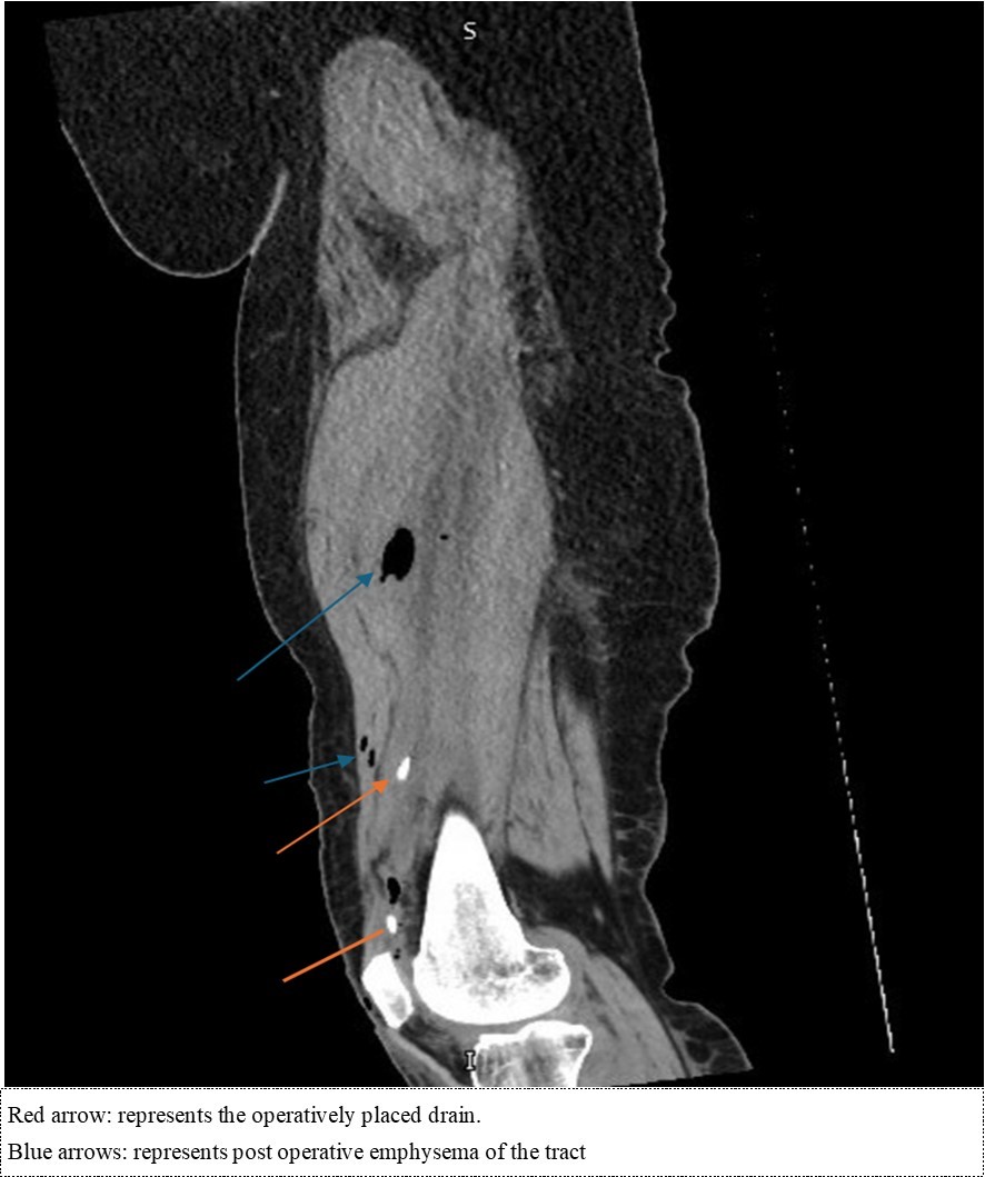
Figure 1b.Coronal cut of the non-contrasted CT femur – soft tissue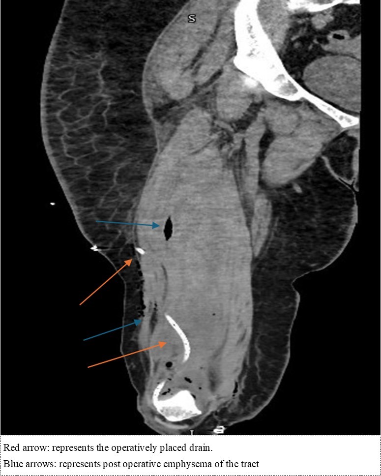
Further history was taken from the patient post-op where she noted that during the 6 weeks when she noted further worsening of her knee pain, she had initially had focal swelling and redness on her right knee with pain on movement. The pain was constant and worsened with movement as well as tenderness. It had progressively worsened with associated progression of the swelling to involve the right knee. She notes 10lbs weight loss and progression of her systemic symptoms. Further history reveals minimal trauma including falls. She also notes that she has 2 pet cats who tend to scratch and bite her on attempts of climbing along her right knee. One of those cats was never vaccinated nor taken to the vet. When asked regarding her knee injection, she un-promptly noted that she felt the IJI needle to unusually track out of her knee and felt it go along the lateral aspect of her distal thigh.
Blood and joint cultures from both the arthrocentesis and intra-op washout were collected. Blood cultures were negative, however, both joint cultures were positive for Streptococcus dysgalactia which was pan-sensitive on MIC susceptibility. The patient’s antibiotic course was de-escalated to amoxicillin 1g three times daily to complete a course duration of 4 weeks. For completion of evaluation, the patient also had a CT of the facial bones to evaluate for causes of hematogenous spread of septic arthritis and was negative. Given the negative blood cultures and negative facial CT, the cause of the septic arthritis was then presumed to be contiguous in nature from untreated cellulitis.
Discussion
As was demonstrated in the case, the patient presented with an unusual clinical case of septic arthritis likely from S. dysgalactiae.This organism is an uncommon cause of skin and soft tissue infections and often occurs in patients with pre-disposing factors such as neoplasms or diabetes mellitus.11 However, in our case, the patient did not have predisposing factors. She likely developed cellulitis from cat bites and scratched introducing the organism, which was not treated, leading to development of contiguously spread septic arthritis.
The knee pain she described was likely due to the ongoing infection, however, due to the chronicity of knee pain in general, was treated as a non-infectious erosive disease such as osteoarthritis. Further history as well as prior ultrasound evaluation may have circumscribed the events that occurred leading to the extra-capsular spread of the disease. Interestingly, the sensation noted by the patient of the needle protruding through the knee and into her lateral thigh was the presumed introduction of the iatrogenic spread of the infection into the thigh.
Looking into the literature, there seems to be benefit of utilization of ultrasound in the evaluation of septic arthritis.12Though operator dependent, it is a readily available tool in sports medicine clinics which are usually used for needle guidance in IJI’s. In the right clinical context, it may be beneficial in evaluation of infectious or non-erosive etiologies prior injection.
Conclusion
Thorough history and physical examination remain as the most important aspects of patient care. They also demonstrate their importance in the initial interview with patients no matter the providers’ specialty in delineating possible etiologies for the patient’s presentation. In arthropathies, following thorough history and physical examination, point-of-care ultrasound are invaluable tools for both diagnostic as well as procedural interventions. This case also attempts to add to the present literature regarding the benefit of ultrasound guidance in both prior to and in the process of joint injections by possibly identifying subcutaneous swelling consistent with skin and soft tissue injection with the potential of reconsideration of the joint injection if concerns for such injections are had.
References
- 1.J S Earwood, T R Walker, Sue G J C. (2021) Septic arthritis: Diagnosis and treatment. American Family Physician.
- 2.Nelson S B. (2024) Septic Arthritis. In:. Current Diagnosis & Treatment: Rheumatology, 4e Stone JH. eds https://accessmedicine.mhmedical.com/content.aspx?bookid=3017§ionid=253717364 .
- 3. (2022) Infectious Diseases: Syndromes and Etiologies. In:. Sherris & Ryan's Medical Microbiology, 8th Edition. McGraw-Hill Education. Accessed October 13 Ryan KJ. eds 2024-3107.
- 4.Cody M E, Tehranzadeh J. (2021) Arthritis and Infection. Basic Musculoskeletal Imaging, 2e. McGraw-Hill Education. Accessed October 13 In: Tehranzadeh J. eds 2024-3075.
- 5.Berger J S, Dangaria H T. (2014) . Joint Injections & Procedures. In: Maitin IB, Cruz E. eds.CURRENT Diagnosis & Treatment: Physical Medicine & Rehabilitation. McGraw-Hill Education. Accessed October 13 2024-1180.
- 6.Foster Z J, Voss T T, Hatch J, Frimodig A. (2015) Corticosteroid Injections for Common Musculoskeletal Conditions. Am Fam Physician. 92(8), 694-699.
- 7.Bookman J S, Pereira D S. (2014) Ultrasound guidance for intra-articular knee and shoulder injections: a review. Bull Hosp Jt Dis. 72(4), 266-270.
- 8.Berkoff D J, Miller L E, Block J E. (2012) Clinical utility of ultrasound guidance for intra-articular knee injections: a review. , Clin Interv Aging 7, 89-95.
- 9.J T Finnoff. (2015) for Sports Medicine (AMSSM) position statement: Interventional Musculoskeletal Ultrasound in sports medicine. , British Journal of Sports Medicine 49.
- 10.In McKean SC, Ross J J, Dressler D D, Scheurer D B. (2017) . eds Principles and Practice of Hospital Medicine, 2e. McGraw-Hill Education. Accessed October 14 2024-1872.