
Use of a Novel Exogenous Allogeneic Multi-Growth Factor Concentrate in Bone Regeneration of the Mandible: A Case Report
Abstract
Regeneration of new bone continues to be one of the most significant challenges in implant dentistry to correct osseous deficiencies in preparation for dental implant surgery. The need for effective bone regenerative strategies has fueled the development of many therapeutic methods, including the use of biologics such as bone morphogenetic proteins, bone marrow aspirates, mesenchymal stem cells, and platelet-derived growth factors. One therapeutic strategy that has not been investigated is the use of exogenous growth factors on bone regeneration of the jaws. The goal of this study is to evaluate if a novel exogenous allogeneic multi-growth factor concentrate, AlloSpark-GF, has a positive effect on bone regeneration in the grafted mandible using histology and histomorphometry.
Article Information
- Received
- Accepted
- Published
Academic Editor: Anubha Bajaj, Consultant Histopathologist, A.B. Diagnostics, Delhi, India.
Checked for plagiarism: Yes
Review by: Single-blind
Copyright © 2026 Cameron Y. S. Lee, et al.
 This is an open-access article distributed under the terms of the Creative Commons Attribution License, which permits unrestricted use, distribution, and reproduction in any medium, provided the original author and source are credited.
Corresponding author: Cameron Y. S. Lee, Private Practice in Oral, Maxillofacial and Reconstructive Surgery. Aiea, Hawaii 96701. The University of Chicago. The Graduate Program. Chicago, IL. 60637 —
Competing Interests
The authors declare no conflicts of interest.
Funding
No specific funding statement was provided by the authors.
Data Availability
No data-availability statement was provided by the authors.
Citation:
Introduction
Dental implant treatment is the standard of care for the restoration of the edentulous jaw to replace missing teeth. Total treatment time can be decreased by surgically placing implants immediately after extraction of teeth. However, this may not be possible due to a deficiency of bone volume in the implant site. Therefore, bone graft augmentation is indicated to correct the osseous deficiency.
In any grafting procedure related to successful implant osseointegration, the objective is the formation of 100% vital bone 1.The ideal graft material is osteogenic, osteoconductive and osteoinductive and can be evaluated by histology and histomorphometric methods to calculate percentages of vital bone formation, residual graft material, and connective tissue 2, 3.
The goal of this study is to evaluate if a novel exogenous allogeneic multi-growth factor concentrate, AlloSpark-GF, has a positive effect on bone regeneration in the grafted mandible using histology and histomorphometry. Prior to initiating this clinical study, the authors conducted an Internet literature search on AlloSpark-GF. There are no published research and clinical studies in the English language literature evaluating the performance of AlloSpark-GF on bone regeneration. Therefore, the benefits of AlloSpark-GF on bone regeneration of the jaws remain unknown.
Materials and Methods
This case study was completed in a private practice (CYSL) and in accordance with the ethical standards of the institutional and national research committee and with the Helsinki Declaration of 1975 that was revised in 2000 4. No ethical approval was required, as the patient’s identity was concealed. Verbal and written informed consent were obtained from the patient to complete the bone graft surgery with extraction of the molar tooth.
A 54-year-old Asian female completed immediate bone grafting after a non-restorable endodontically treated molar tooth was extracted in preparation for future implant surgery. Two bottles of lyophilized AlloSpark-GF (0.5 cc) allograft powder are hydrated with 2.0 cc of sterile water and incorporated with one gram of cortical allogeneic bone (Salvin Dental). According to the manufacturer, AlloSpark-GF contains high concentrations of growth factors which include the following: bone morphogenetic protein (BMP 2, 4, 6, 7, 9), transforming growth factor beta (TGF-b), vascular endothelial growth factor (VEGF), platelet-derived growth factor (PDGF-BB), and epidermal growth factor (EGF). For the anxious patient, an advantage with use of this exogenous multi-growth factor concentrate is avoidance of the phlebotomy procedure to obtain venous blood required with PRP and PRF to obtain autologous growth factors.
The graft material was passively packed into the extraction site after the molar tooth was extracted and the mandible debrided. A non-resorbable membrane was placed directly over the graft material to prevent epithelial migration into the graft site and removed at the time of implant surgery. After 103 days of bone graft healing time, using a trephine drill with a 2.0 mm internal diameter and length of 10.0 mm was used to harvest the bone core from the posterior mandible. The bone core sample was placed in 10% buffered formalin and submitted for histologic examination at the Hard Tissue Laboratory of the University of Minnesota School of Dentistry.
Results
A single bone core was obtained from the #30 molar site at the time of implant surgery. Vital new bone formation was evaluated using histologic and histomorphometric analysis at 103 days post-bone grafting. Histological analysis revealed direct contact of allogeneic bone with newly formed autogenous bone with continuous osteoid formation. The presence of osteocytes in their lacunae was identified as vital bone compared to residual graft material that did not contain osteocytes. Histomorphometry demonstrated 87.3% vital new bone. Images of de novo bone formation are shown in Figure 1, Figure 2, Figure 3, Figure 4, Figure 5.
Figure 1. Medium power histology view. Extensive formation of mature vital bone. Note bridging between sheets of vital bone. VB: mature vital bone. AL: Allogeneic bone. MS: marrow space. NB: immature new bone. Original magnification x 40. Stevenel’s blue and Van Gieson’s picro fuchsin.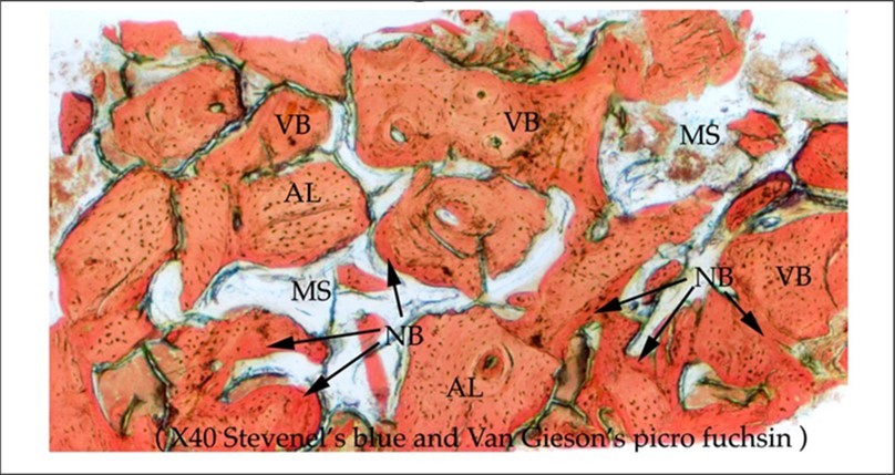
Download figure
Figure 2. Medium power histology view. Formation of immature new bone with osteoid formation. VB: mature vital bone. AL: Allogeneic bone. NB: immature new bone. MS: Marrow space. OD: Osteoid. Original magnification x 40. Stevenel’s blue and Van Gieson’s picro fuchsin.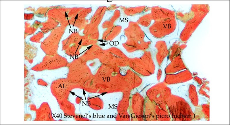
Download figure
Figure 3. High power histology view. Neovascularization illustrated by the presence of multiple haversian canals that provide the vascular supply to newly formed osteocytes. VB: mature vital bone. NB: immature new bone. MS: Marrow space. HC: Haversian canal. FM: Fibrous marrow. Original magnification x 100. Stevenel’s blue and Van Gieson’s picro fuchsin.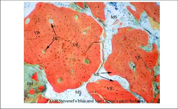
Download figure
Figure 4. High power histology view. Dense new vital autogenous bone with a wide seam of green-staining osteoid. VB: mature vital bone. NB: immature new bone. MS: Marrow space. HC: Haversian canal. FM: Fibrous marrow. OD: Osteoid. Original magnification x 200. Stevenel’s blue and Van Gieson’s picro fuchsin.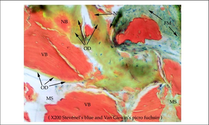
Download figure
Figure 5. High power histology view. Extensive formation of immature new bone with mature vital bone. Note the numerous osteocytes in their lacunae and osteoblasts all involved in osteogenesis. NB: immature new bone. OD: Osteoid. Original magnification x 200. Stevenel’s blue and Van Gieson’s picro fuchsin. 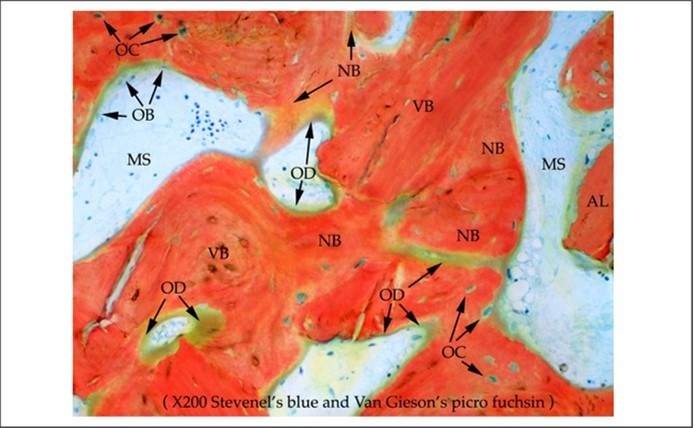
Download figure
Discussion
Use of growth factors obtained from platelet rich plasma (PRP) and platelet rich fibrin (PRF) obtained from autologous blood of the patient has successfully been used in hard and soft tissue wound healing 5, 6, 7. Platelets contain osteoinductive growth factors that stimulate bone formation by directly promoting the osteogenic lineage targeting osteoblasts such as bone morphogenetic protein (BMP), transforming growth factor beta (TGF-b), vascular endothelial growth factor (VEGF), platelet-derived growth factor (PDGF), and epidermal growth factor (EGF). These growth factors accelerate the wound healing process and tissue regeneration by accelerating cell division, proliferation, differentiation and promoting the migration of stem cells 8, 9, 10, 11.
In clinical studies with mandibular reconstruction, Marx et al demonstrated that the addition of PRP resulted in early graft consolidation and mineralization in half the time compared with grafts without the addition of PRP 12. Sohn and colleagues reported that in maxillary sinus augmentation and alveolar bone grafting, concentrated growth factors were effective in neovascularization and new bone formation 13. Therefore, efforts have been made to use concentrated growth factors obtained from platelets in autologous blood of the patient that do not cause infection and hypersensitivity reactions, have a simple preparation, and are less invasive 7.
AlloSpark-GF (Salvin Dental/Young Innovations) contains over 1,000 naturally derived proteins, growth factors, and peptides. High concentrations of growth factors include the following: bone morphogenetic protein (BMP 2, 4, 6, 7, 9), transforming growth factor beta (TGF-b), vascular endothelial growth factor (VEGF), platelet-derived growth factor (PDGF-BB), and epidermal growth factor (EGF). The histological results of the present study showed that AlloSpark-GF accelerated bone regeneration over a three-month period (Figure 1, Figure 2, Figure 3, Figure 4, Figure 5).
Bridging of sheets of residual allograft with newly formed autogenous bone is observed in Figure 1 and Figure 2. Neovascularization is actively occurring as multiple haversian canals that provide the vascular supply during osteogenesis are observed (Figure 3). Note the presence of green colored staining osteoid secreted by osteoblasts that will transform into mature lamellar bone (Figure 4 and Figure 5). Numerous osteocytes are observed that regulate the cell cycle derived from osteoblasts (Figure 5).
Oliveira et al (2021) showed that use of multiple growth factors has a greater effect on bone regeneration compared to use of only a single growth factor. The authors of this study hypothesize that early bone formation and the high percentage of new vital bone are attributed to the enhanced and prolonged healing effects of the multiple growth factors. The rich source of growth factors may function as a chemoattractant for cells at the surgical site, such as neutrophils, fibroblasts and macrophages that are all involved in soft and hard tissue healing.
Of the different growth factors in AlloSpark-GF, the authors believe that VEGF is one of the most important growth factors in bone formation as VEGF not only regulates osteogenesis and angiogenesis, but cell recruitment and cell differentiation.
Conclusion
Bone regeneration is an area of intense scientific and clinical research in implant dentistry. To accelerate bone regeneration, therapeutic strategies include the use of biologics to accelerate bone maturation and bone formation. This is the first clinical study that demonstrates that AlloSpark-GF can accelerate bone regeneration over a three-month period. This case report does have limitations. As this is a single case report, the authors do not know the full potential of this multi-growth factor concentrate on bone graft regeneration. This is one of two ongoing research studies with a cohort of patients that will be presented. A second clinical study is in progress that will compare platelet derived growth factors with Allospark-GF on bone regeneration.
Disclosure
The authors received material support from Salvin Dental and Young Innovations to conduct this study.
References
- 1.Garetto L P, Chen J, Parr J A. (1995) Remodeling dynamics of bone supporting rigidly fixed titanium implants: A histomorphometric comparison in four species including humans. Implant Dent 4, 235-243.
- 2.Block M S, Kent J N. (1997) Sinus augmentation for dental implants: The use of autogenous bone. J Oral Maxillofac Surg 55, 1281-1286.
- 3.Del Fabbro M, Testori T, Francetti L. (2004) Systematic review of survival rates for implants placed in the grafted maxillary sinus. Int J Periodontics Restorative Dent. 24, 565-578.
- 4.World Medical (2000) Association Declaration of Helsinki Ethical Principles for Medical Research Involving Human Subjects. World Medical Association Declaration of Helsinki: Ethical principles for medical research involving human subjects. JAMA. 284-3043.
- 5.Marx R E. (2004) Platelet rich-plasma: Evidence to support its use. J Oral Maxillofac Surg 62, 489-496.
- 6.Anitua E, Andia I, Ardanza B. (2004) Autologous platelets as a source of proteins for healing and tissue regeneration. Thromb Haemost 91, 4-15.
- 7.Dohan D M, Choukroun J, Diss A. (2006) Platelet-rich fibrin (PRF): A second-generation platelet concentrate. Part I: Technological concepts and evolution. Oral Surg Oral Med Oral Pathol Oral Radiol Endod 101-37.
- 8.Intini G. (2009) The use of platelet-rich plasma in bone reconstruction therapy. Biomaterials 30, 4956-4966.
- 9.Amini A R, Laurencin C T, Nukavarapu S P. (2012) Bone tissue engineering: recent advances and challenges. Crit Rev Biomed Eng [Internet]. 40(5), 363-408.
- 10.Cui Q, Dighe A S, Irvine Jr JN. (2013) Combined Angiogenic and Osteogenic Factor Delivery for Bone Regenerative Engineering. Curr Pharm Des [Internet]. 3374-83.
- 11.Ramasamy S K, Kusumbe A P, Itkin T, Gur-Cohen S, Lapidot T et al. (2016) Regulation of Hematopoiesis and Osteogenesis by Blood Vessel–Derived Signals. Annu Rev Cell Dev Biol 32(1), 649-75.
- 12.Marx R E, Carlson E R, Eichstaedt R M. (1998) Platelet-rich plasma: Growth factor enhancement for bone grafts. Oral Surg Oral Med Oral Pathol Oral Radiol Endod. 85, 638-646.
- 13.Sohn D S, Moon J W, Moon Y S. (2009) The use of concentrated growth factors (CGF) for sinus augmentation. J Oral Implant. 38, 25-38.
- 14.Dragonas P, Schiavo J H, Avila-Ortiz G. (2019) Plasma rich in growth factors (PRGF) in intraoral bone grafting procedures: A systematic review. J Craniomaxillofac Surg. 47, 443-453.
- 15.Oliveira E R, Nie L, Postawczyk D. (2021) Advances in growth factor delivery for bone tissue engineering. Int J Mol Sci 22, 903-907.